- Visibility 92 Views
- Downloads 7 Downloads
- DOI 10.18231/j.ijogr.2020.131
-
CrossMark

Open myomectomy as an approach to removal of multiple large uterine fibroids
Case 1
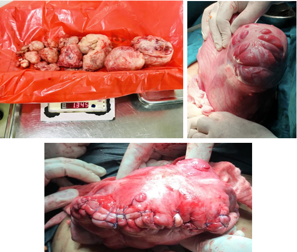
A 32yr old female, nulligravida married since 4 years, eager to conceive presented to us with complaints of heavy menstrual bleeding and a large abdominal mass. Patient had a history of multiple blood transfusions previously due to anemia occurring as a result of heavy menstrual bleeding.
On examination her uterus was around 28 weeks size hard firm. Ultrasound of the abdomen was done which was suggestive of multiple fibroids with degenerative changes the largest being 10*9cm.
Patient was admitted for an open Myomectomy after reserving adequate blood and need for SOS hysterectomy. Intraoperatively the uterus was enlarged with multiple uterine fibroid of varying size and with degenerative changes. 15 fibroids were removed weighing a total of 1.345kg. Patient required multiple blood transfusions however was discharged on post-operative day 5.
Case 2
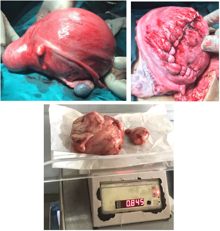
A 35-year old nulligravida married for 2 years eager to conceive presented to us with complaints of pain in abdomen on and off since 2-3 years.
On examination per abdomen her uterus was around 24 weeks size, firm, mobile.
Ultrasound of abdomen and pelvis was done s/o- bulky uterus with multiple fibroids, largest in fundal region measuring 13.92*11.94 cm.
Patient was taken for an open Myomectomy. A large fund fibroid of around 12cm and 2 small fibroids from the posterolateral wall were removed weighing a total of 845gms.
Patient was discharged on POD5.
Patient followed up at regular intervals 1 month, 3 months, 6 months and 1 year.
Case 3
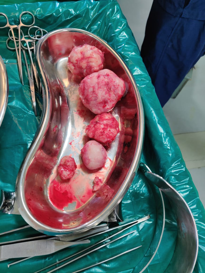
A 32-year-old female X, Parity1 abortion 1 previous LSCS presented to us with complaints of pain in abdomen on and off since 3 years. On examination the uterus was 20-22weeks. On ultrasound, uterus size was 12.2 x 7.8 x 10.4 cm anteverted, bulky uterus. Multiple heteroechoic predominant hypoechoic with evidence of minimal vascularity suggestive of anterior wall fibroid 4.6 x 5.3 cm in upper segment and 3.9 x 3.9 cm in lower segment, posterior wall fibroid 3.7 x 4.5 cm, pedunculated fibroid 9.9 x 6.8 cm arising from uterus extending to left adnexa. On CT scan revealed a large fibroid 19 x 11 x 10 cm fibroid.
Open myomectomy done, largest fibroid removed from anterior wall 16 x 8 cm.
Material and Methods
Preoperative preparation: All patients were kept Nil by mouth for 12 hrs and adequate bowel preparation was given. Adequate blood and blood products were reserved preoperatively. Average duration of operative procedure was 1and ½ to 2 hrs.
Operative procedure
Intraoperatively Foley’s catheter was kept in uterine cavity and infiltrated with 30cc normal saline + methylene blue. This would help to diagnose if uterine cavity was accidently opened.
Abdomen was opened in layers, uterus was exteriorized and infiltration was done with diluted vasopressin. Myoma was hooked with myoma screw and single uterine incision was made. Myoma was separated from the capsule, base clamped and hemostatic sutures taken.
Uterine cavity was not opened
Intraoperative blood loss was managed by blood and blood product transfusion.
Postoperative anemia was corrected by blood transfusion.
Patients were discharged on post-operative day 5. Patients were followed up at intervals of 1 month and 3 months, 6 months and 1 year.
Patients were assessed on the basis of post operative pain, menstrual regularity, fertility and any abnormal uterine bleeding.
Results
On follow up assessment all three patients achieved regular menstrual cycles after 3 months interval from the operation. Hysteroscopic evaluation for adhesions was not done however none of the patients reported abnormal uterine bleeding. The degree of post operative pain at intervals of 1month was 5/10 and 0/10 on subsequent follow up visits. One out of the 3 patients had a spontaneous conception 6 months post surgery.
Discussion
Uterine fibroids are the most common gynecological tumors occurring in women in their reproductive years. Fibroids can lead to various complications. These are symptomatic in around 50% women. Myomectomy is defined as surgical removal of uterine fibroid or myoma There are various modalities available for management of fibroids including conservative oral and parenteral treatment, conservative and radical surgery, and newer techniques such as uterine artery embolization and radiofrequency ablation.[3]
Given their size and location, it is unsurprising that simple physical impedance to the transport of sperm, egg or embryo has been proffered as a mechanism to explain the anti-fertility effects of fibroids. Location rather than size of fibroid is of importance submucous myomas distorting the uterine cavity are particularly implicated in infertility.[4]
The process of describing the location, size, and character of uterine fibroids, commonly referred to as ‘fibroid mapping’.Myoma mapping is an important component of planning any procedure for myoma removal. It is a definite and reproducible method to describe the entire disease. Earlier crude methods such as physical examination and hystero-salpingography were used however with modern advancement many new techniques are available. A 2 dimensional ultrasound is the most commonly available and used, a transvaginal scan is considered superior to a trans- abdominal scan. Other modalities such as Three Dimensional Ultrasonography, Infusion Sonohysterography, 3D Sonohysterography, Magnetic Resonance Imaging (MRI), Conventional Hysterosalpingography (HSG) and Computed Tomography Scan (CT scan) are also now being used.[5]
The method preferred in this particular study was Open myomectomy. The most common complication of open myomectomy is intraoperative haemorrhage requiring blood transfusion. In a study conducted by Kikelomo T Adesina et al. the incidence was found to be 21.2%.[1] The use of vasopressin infiltration has shown to reduce the incidence of intraoperative bleeding, in a study conducted by H. Fletcher et al. there was <800ml blood loss in vasopressin group compared to 1000ml in tourniquet group.[6]
Another complication associated with open or laparoscopic myomectomy is breach of endometrial cavity after inability to determine the correct plane and in case of smaller fibroids closer to the uterine cavity.[7] In a study conducted by Gupta et al. 2013 there was a 30% incidence of post-operative adhesions following breach of endometrial cavity.[8]
The use of an intrauterine balloon post-operatively has demonstrated to reduce the incidence of post-operative adhesions. In a pilot study conducted by Dr. Sadhana Gupta et al. it was found that in cases where there was breach of the uterine cavity during myomectomy insertion of a intrauterine balloon till 4th postoperative day reduced the incidence of postoperative adhesions. There were intrauterine adhesions in 3 out of 10 women in control group compared to no adhesions in 16 women in study group on 6month follow up hysteroscopy.[9]
Intrauterine balloon can also be place preoperatively.[10] This offers the following advantages:
Palpation of the Foley catheter balloon through the myometrium can provide the surgeon with information about the location of the cavity.
The Foley catheter can be used to inject methylene blue into the uterine cavity, aiding diagnosis of cavity breach.
Repair of the endometrium is facilitated by the presence of the Foley balloon.
If a cavity breach occurs following repair of the defect, the Foley balloon can be left in the uterine cavity for 10 days to minimize the risk of intrauterine adhesion formation[11]
Laparoscopic myomectomy is the preferred surgical management of fibroids however it has the disadvantage of longer operating time, specialized equipment and surgical expertise.
Open myomectomy offers the advantage of removal of all myomas through a single uterine incision with tunneling as opposed to laparoscopic surgery which requires multiple uterine incisions and also a shorter operating time and hence was the modality opted for in this case report.
In a meta-analysis conducted by Jin et al(2009) comparing laparoscopic and open myomectomy for patients with fibroids laparoscopic myomectomy was associated with decreased blood loss during surgery. Data showed that postoperative pain was also diminished. However, it took longer operation time than abdominal myomectomy. Other parameters such as rate of recurrence of myoma and rate of postoperative pregnancy were similar in both LM and OM groups.[12]
However, because of ease of surgery and lack of equipment open myomectomy for large uterine fibroids is still the procedure of choice in many centres worldwide.[6]
The aim of this case study was to demonstrate that open myomectomy still has a role in modern day gynaecological surgery. If performed with adequate preoperative preparation and skilled surgeon can lead to good patient outcome comparable to laparoscopic myomectomy.
Source of Funding
None.
Conflict of Interest
The authors declare that there is no conflict of interest.
References
- K T Adesina, B O Owolabi, H O Raji, A O Olarinoye. Abdominal myomectomy: A retrospective review of determinants and outcomes of complications at the University of Ilorin Teaching Hospital, Ilorin, Nigeria. Malawi Med J 2017. [Google Scholar] [Crossref]
- L Mettler, T Schollmeyer, A Tinelli, A Malvasi, I Alkatout. Complications of Uterine Fibroids and Their Management, Surgical Management of Fibroids, Laparoscopy and Hysteroscopy versus Hysterectomy, Haemorrhage, Adhesions, and Complications. Obstet Gynecol Int 2012. [Google Scholar] [Crossref]
- H Fletcher, C Burrell, B Bassaw, E Thomas. Complications of Uterine Fibroids and Their Management. Obstet Gynecol Int 2012. [Google Scholar] [Crossref]
- P. Purohit, K. Vigneswaran. Fibroids and Infertility. Curr Obstet Gynecol Rep 2016. [Google Scholar] [Crossref]
- B Nusair, M Al-Gudah, R Chodankar, I A Abdelazim, M Abu Faza. Uterine Fibroid Mapping. Curr Obstet Gynecol Rep 2016. [Google Scholar] [Crossref]
- H Fletche, J Frederick, M Hardie, D Simeon. A Randomized Comparison of Vasopressin and Tourniquet as Hemostatic Agents During Myomectomy. Obstet Gynecol 1996. [Google Scholar] [Crossref]
- K Rajah, M Dizdar, N Balachandren, K Kriedt, E Saridogan, D Mavrelos. Who is at risk of endometrial cavity breach at laparoscopic myomectomy?. Facts Views Vis Obgyn 2019. [Google Scholar]
- S Gupta, V S Talaulikar, J Onwude, I Manyonda. A pilot study of Foley’s catheter balloon for prevention of intrauterine adhesions following breach of uterine cavity in complex myoma surgery. Arch Gynecol Obstet 2013. [Google Scholar] [Crossref]
- S Gupta, V S Talaulikar, J Onwude, I Manyonda. A pilot study of Foley’s catheter balloon for prevention of intrauterine adhesions following breach of uterine cavity in complex myoma surgery. Arch Gynecol Obstet 2013. [Google Scholar] [Crossref]
- H H Hatasaka, D T Carrell, C M Peterson. Acquired uterine factors and infertility. Reproductive Endocrinology and Infertility 2010. [Google Scholar]
- M Afnan, A Coomarasamy. Myomectomy: Breach of the Endometrial Cavity. Gynecologic and Obstetric Surgery 2016. [Google Scholar] [Crossref]
- C Jin. Laparoscopic versus Open Myomectomy-A Meta-Analysis of Randomized Controlled Trials. Eur J Obstet Gynecol Reprod Biol 2009. [Google Scholar]